19.06.2026
BSMU scientists develop IT solutions for medicine using artificial intelligence
Artificial intelligence helps automate work not only with documentation. In medicine, it helps in the interpretation of X-rays, MRI and other images, is able to detect signs of cancer and fractures with an accuracy inaccessible to the naked eye.
Scientists of the Bashkir State Medical University of the Ministry of Health of Russia are actively working to develop programs with the safe use of artificial intelligence in education and medicine.
"New information technologies, IT technologies, digital technologies that we are introducing today into the work of our University are not a tribute to fashion, but an urgent need, an element of the development of a modern University," he rector of the Bashkir State Medical University of the Ministry of Health of Russia, Academician of the Russian Academy of Sciences, Professor Valentin Pavlov said.
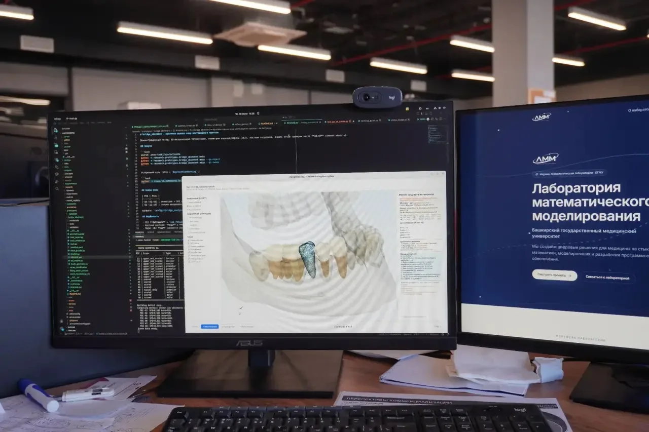
Thus, scientists from the laboratory of mathematical modeling of BSMU at the site of the Interuniversity Student Campus are developing a "dental assistant" - a program that will help the dentist analyze three-dimensional images of teeth and jaws. Such images contain a large amount of information and help to assess the condition of teeth, roots, bone structures and other areas to the smallest detail. The program will enable the doctor to quickly identify clinically significant areas for treatment, the system visualizes the teeth in a three-dimensional image, which makes it possible to better assess the degree of bone tissue damage.
For example, there is currently a high-precision 3D-KLKT X-ray diagnostic method (cone beam computed tomography) that creates a 3D image of the dentoalveolar system. In practice, such a snapshot is a large amount of data that needs to be processed based on professional experience. An AI-based program developed by BSMU scientists concentrates all the necessary data in a single space. The system quickly displays a ready-made anatomical model and notes important features for the doctor: changes in the shape, position, condition of the roots, bone support, as well as a defect in the dentition. This speeds up the preparation of the conclusion and treatment plan. The solution is applicable both in the clinic and in the training of students, residents and novice doctors.
Currently, the BSMU of the Ministry of Health of Russia is implementing large-scale scientific programs for the development of robotic surgery. Recall that 8 years ago, the first robotic surgery center in the Volga Federal District was opened at the BSMU Clinic. The founder and ideological inspirer of the center is the rector of the Bashkir State Medical University of the Ministry of Health of Russia, academician of the Russian Academy of Sciences, Professor Valentin Nikolaevich Pavlov. Currently, the Center has 3 robotic installations, including a robotic joint replacement traumatologist - Cuvis Joint. In 2025, the Center performed more than 1,000 interventions on two Da Vinci robots and about 300 on a trauma robot. The applications of Da Vinci technology are very extensive and include abdominal, thoracic and colorectal surgery, gynecology and urology, oncology and vascular surgery.
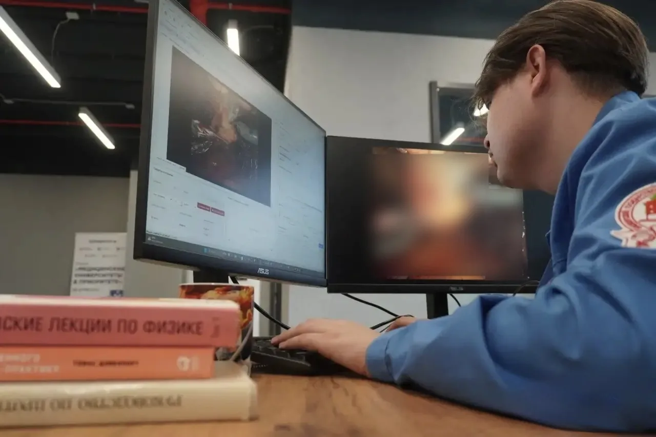
Now scientists are working on the development of a program using AI - "OpeLab." The system will help analyze video of robot-assisted operations performed using the Da Vinci Xi robotic surgical system. The task of the program is to turn video into specific data based on various stages of operations: the tools used, the time they appear in the frame, the trajectory of movement, the path traveled, the speed of movement, the duration of the stages of the operation, periods of activity and pauses, the synchronicity of the tools. The knowledge gained will become the basis for an objective analysis of operations in order to train surgeons, analyze the technical features of the operation, scientific research and further the formation of educational materials. Note that the program does not "evaluate" the surgeon, but provides measurable parameters for expert analysis.
Scientists of the Bashkir State Medical University of the Ministry of Health of Russia contribute to the prevention and prevention of diseases of the cardiovascular system using AI. Heart and vascular diseases consistently remain the leading cause of death worldwide. For the analysis of angiographic images of coronary vessels, specialists from the mathematical modeling laboratory are developing the ArtPiece program.
* Coronary angiography is the main method of diagnosing heart disease, in which a safe contrast agent is injected into the vessels and a series of X-rays is taken. This allows the cardiologist to see the exact locations of the narrowing of the arteries and choose the most effective treatment.
The AI-based program will make it possible to qualitatively supplement the images and visual assessment of a specialist doctor, assess in more detail the severity of the coronary bed lesion and then choose the optimal treatment method. For example, indicators are calculated after highlighting the contour of the vessel and plotting its central line with an estimate of the length of the segment, the diameter/width of the gap in the image area, the degree of change in diameter, indicators of crimp, curvature, bending angles, the ratio of the actual length of the vessel to the rectilinear distance. The program selects vessels, analyzes their shape, calculates crimp and finds areas that require additional attention, and then generates a report. Automatic isolation of the vascular bed saves time and reduces the dependence of the result on manual marking. The knowledge gained helps to understand and predict the behavior of biological processes, the dynamics of diseases and the effectiveness of treatment. This allows the development of new methods of diagnosis and therapy, as well as the optimization of existing treatment approaches.